Features
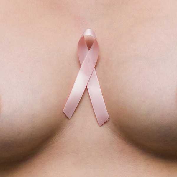
BN Doctors’ Lounge: Pink October – Breast Lumps
 One of the most alarming breast symptoms reported by women is breast lumps and understandably so. Breast cancer is the most invasive cancer in women and accounts for over half a million deaths every year worldwide. Today, we will look in detail at lumps in the breasts. What they are, how many types there are, which ones are cancerous and what one can do to treat them.
Next week we will look at nipple discharge, skin conditions affecting the breast and other breast diseases.
I support a charity called BRECAN which stands for Breast Cancer Association of Nigeria, and this year for our Pink October breast Cancer Awareness program, the wonderful people at SHOPRITE have given us a ₦20,000 gift voucher to give away. All you need to do to win is take a creative picture or video of you wearing something pink, add a message and share it with us on Facebook, Twitter, or Instagram, by using the hashtags #PinkNigeria #BrecanCares. I’ll be looking out for your pictures, so get snappy.
There’s a Lump in My Breast! Do I Have Cancer?
There are different causes of breast lumps and unlike breast pain, it is not as easy to determine if a lump in the breast is a cause for concern or not. Ultimately, the all clear can only be given after a biopsy -removing some or the entire lump, and looking at the cells it’s made up of under the microscope, has been done. There are several conditions that can have a lump as part of their features and these include the following:
• Fat necrosis
• Cysts
• Fibroadenosis
• Fibroadenoma
• Cancer
A few other conditions already described in previous articles could also be present as a lump. For instance, the lumpiness in cyclical mastalgia may mimic a breast lump and a breast abscess may also be felt as a lump in the breast.
NON CANCEROUS LUMPS
Fat necrosis
The breasts are made up of a significant amount of fat. When there is blunt trauma to the breasts the fat cells can get injured. The resulting injury triggers a change in the consistency of the area from soft to hard and this can often be felt as a lump under the skin. Fat necrosis can also be mistaken for cancer because it often gets calcified (crystals of calcium get deposited on its surface) and calcification is also seen in some types of breast cancer. Fat necrosis is harmless however and is no cause for alarm.
Cysts
Cysts are fluid filled sacs, and can form within the glands of the breast. It is felt as a squishy compressible lump in the breast usually just under the areola or further out in the breast. The fluid contained in the sac will determine what kind of cyst it is. There could be blood filled cysts, milk filled cysts and clear fluid filled cysts. Most cysts are harmless and your doctor will take a sample of the fluid with a syringe to determine what kind of cells are in it (fine needle aspiration cytology)
Some cysts may be pre-malignant and we will discuss these next week.
Fibroadenosis
These are also called Lumpy Lumps. Remember the lumpiness we said often occurs during the beginning of a woman’s menstrual period? This lumpiness is usually indistinct and ill defined and it is often not possible to say where the lump ends and normal breast tissue begins. Sometimes however two or more of these may fuse together, to form a distinct lump that can be felt as a solid lumpy mass within the breast. Usually, no treatment is required and most doctors will want to leave lumps of this sort alone as most cases of fibroadenosis will resolve on their own. In some instances however, the patient may require an excision biopsy (lumpectomy) and this will be discussed in a later section.
Fibroadenoma
Also called the breast mouse, these lumps are small, discreet and very mobile, just like a mouse. (Mobile here means that the location of the lump can seem to vary greatly and is often said to feel as though the lump is ‘running around’ the breast when you feel for it). As with a fibroadenosis, fibroadenomas are benign (are not cancerous) and can be left alone. However, in some instances like family history of breast cancer or recurrent fibroadenomas in the past, your doctor may decide to do an excision biopsy (lumpectomy) to ascertain the cell types in the lump.
Breast Cancer
A cancerous breast lump is often described as hard, irregular, and fixed meaning that it is not freely mobile within the breast. It can occur in any part of the breast but is often found in the upper outer side of the breast, near the armpit. Cancerous breast lumps usually start out painless and only become painful after the disease has significantly progressed. It can vary in size from small to large and will typically get bigger in size as the disease progresses.
Other signs of breast cancer include dimpling of the skin, nipple retraction, bloody nipple discharge and recent breast anisometry.
What Causes Breast Cancer?
The human body is made up of trillions of cells each of which have a specified life span. During the life of a healthy cell, it will multiply by dividing itself a number of times to produce daughter cells that will take over its function when it dies. This cycle of cell division and normal cell death is tightly regulated by specialized genes in our DNA. In normal conditions, only healthy cells are allowed to multiply and produce daughter cells and this prevents the multiplication of damaged cells which would be unable to function adequately.
Cell damage is caused by charged particles called free radicals which are produced by ionising radiation like x-rays, chemicals like benzene, some viruses and certain foods. Once these free radicals get into the body they are capable of irreversibly damaging the DNA of exposed cells and once this occurs, the deformed cell is incapable of performing its normal function. The body counters this cell damage by an Antioxidant clearance system which prevents the formation of free radicals and minimises their effect as well as a sentinel system known as Apoptosis which identifies damaged cells and ensures that they are all killed and removed from the body.
Sometimes however, this double security layer can fail if circulating levels of antioxidants are not adequate enough to prevent free radical formation and if the resulting damaged cell escapes apoptosis. If this damaged cell begins to replicate itself and its daughter cells also divide and produce more damaged cells, the resulting mass of unhealthy cells is what is known as a cancer. Healthy cells have programmed within their DNA a specific number of times that they are allowed to replicate and this ensures that tissues and organs retain a particular shape and size. Damaged cells on the other hand have lost the replication limit control and this means that a group of cancer cells can go on replicating as often as possible and can increase in size indefinitely.
One of the cardinal causes of cancer is the failure of the sentinel system of apoptosis and this system failure is more likely to occur in cells that grow rapidly and have a rapid turnover of cells. It’s almost as though there are only a few customs officers looking out for suspected smugglers at a very busy border post. Even if each officer worked all day, there would still be more contraband items entering through this post than would have if it were a less busy border. This is one of the reasons that the breast is prone to developing cancer because as soon as puberty begins until menopause, the breast tissue is under a perpetual cycle of accelerated growth followed by accelerated regression driven by the female sex hormones. This rapid cell growth and regression makes it more likely for damaged cells to escape detection and by so doing replicate to form a cancerous growth.
Also, certain families have a gene that can make having breast cancer more likely and these genes are the BRCA1 and BRCA2 genes. A woman who inherits this gene has a higher chance of coming down with breast cancer. Gene testing for the BRCA1&2 genes are available. If you have had more than three members of your family who have had breast cancer, it may be beneficial to find out your BRCA status.
How Will the Doctor Know if I Have Breast Cancer?
After listening to your symptoms and asking about family history, your doctor will examine your breasts and feel for the lump to characterize it. Depending on your age, your doctor will have you do either a breast ultrasound or a mammogram to visualise the lump and surrounding tissue. Some lumps are fluid filled and your doctor will want to take a sample of the fluid with a fine needle and send it to the lab to determine what cells are in it. For solid lumps, your doctor could either take a piece of the lump or send for biopsy, (tru-cut™core biopsy) or schedule you for a surgery called an excision biopsy or lumpectomy to remove the entire lump and send it for analysis.
Lumpectomy and Excision Biopsy
If a lump is suspicious, it is current best practice to remove it and send it to the lab for testing to determine its cellular constituents. All suspected cancerous lumps should be removed and tested, while fibroadenomas and fibroadenoses may or may not be removed.
Most lumpectomies are done under general anaesthesia and may are may not include axilliary clearance (removal of armpit lymph nodes).
Just before your surgery, your surgeon may make some marks on the surface of the breasts with a surgical marker to help define the exact location of the lump and an anaesthesiologist will put up an intravenous line and will give you a relaxing medication as well as numbing injections to the breasts.
The surgery begins with a small curved incision along the natural curve of the breast to expose the lump and once it is visualised or felt in its entirety, the surgeon will remove the whole lump as well as a chunk of surrounding tissue. Sometimes a rubber tube called a surgical drain is put into the wound to drain any blood that pools in the space once occupied by the lump. The wound is then closed with either stitches or staples but most surgeons would use a fine absorbable suture that will dissolve over time and leave minimal scarring. Typically your operation should last between 15-30 minutes.
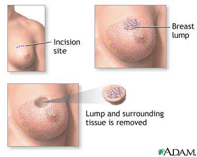
After the procedure, you will be wheeled into a recovery room and your vital signs monitored until you are stable. Lumpectomies are usually day cases and overnight stay is often not necessary.
Pain medication will be prescribed and your doctor will advise you what signs of infection to watch out for and when to return for a follow up visit to inspect the wound and remove stitches or staples if need be.
At home, you will need to keep the wound area clean and dry and keep the breast supported. You may find it more comfortable to sleep face up or on the opposite side of the operation site. Get lots of rest, eat well, drink lots of fluids and exercise your arm to prevent stiffness.
What Next?
After your lumpectomy or fine needle aspiration, the tissue samples are sent to the lab for analysis and after a few days or a week, your results should be ready for you to discuss with your doctor. Almost all fibroadenomas and fibroadenoses will come back benign and your doctor will give you an all clear. In women with suspicious lumps, some will receive the news that the lump removed was actually cancerous and if this is the case, your doctor will discuss with you what treatment is best suited for the particular stage of the cancer and what adjuvant therapies may be necessary. In some women, the lumpectomy may be almost all that was necessary to treat the disease as the cancer would have already been removed. All that remains to complete treatment would be radiation or chemotherapy or hormonal treatment depending on what the cancer is sensitive to. In most women however, it may be necessary to remove the entire breast as well as surrounding tissue and lymph nodes in a procedure called a mastectomy to completely remove the cancer. After a mastectomy, women usually receive chemotherapy or radiotherapy or both. Oestrogen receptor positive cancers will also benefit from hormonal therapy.
If you have felt a lump in your breast, please see a physician.
Can I Prevent This?
Breast cancer, like all cancers is caused by DNA damage to cells and theoretically, the best way to prevent cancer is to prevent cell damage. However, our modern lifestyle puts us in contact with a vast number of potential carcinogens on a daily basis and if you read the news, then you may have concluded that almost everything is cancerous these days. From second hand cigarette smoke to plastic bottles releasing xeno-oestrogens to free radicals in Suya and aflatoxins in groundnuts and Amala, it seems like there is no escaping these cancer causing substances.
The organic movement in the US, UK and Canada encourages people to shun processed foods and chemicals and return to consuming only natural products. This is even more attainable here in Nigeria as most of our vegetables, poultry and agricultural produce are farm grown, free range and without harmful chemicals.
Eating foods rich in antioxidants is also very helpful as this will prevent oxidative damage to cells and minimise the effect of free radicals in the system.
Eating healthy foods, drinking lots of water and exercising daily can also help keep the immune system healthy.
Finally, stop smoking, maintain a healthy weight, cut down on alcohol consumption, avoid known carcinogens and check your breasts regularly to ensure early detection.
In a Nutshell
There are two types of breast lumps, Non cancerous and cancerous. Majority of lumps found in young women are benign and require no treatment. A hand full of women will have a positive cancer diagnosis and will require some sort of additional treatment including possible mastectomy. Breast cancer is beatable and there a many survivors. The key to survival is early detection and prompt treatment. Please check your breasts today.
Did You Know?
Some women with a positive BRCA gene may opt for a pre-emptive double mastectomy to remove both breasts before they develop cancer and famous Hollywood actress, Angelina Jolie is said to have had the procedure. The breast tissue may be replaced by saline implants.
Disclaimer: This article is written for patient education. It is not intended to diagnose or prescribe treatment and does not replace the advice of your physician. It in no means attempts to cover the full medical scope of this condition. All images are the copyright of their respective owners and are used for illustrative purposes only.
Photo Credit: ©A.D.A.M medical images, All rights reserved | ©Bingeandpurge . All rights reserved.
_______________________________________________________________________________
David Olamide Craig is a Physician, Pastor, Photographer, Poet and Playwright. He graduated Bachelor of Medicine and Bachelor of Surgery from the University of Ibadan and holds a Masters in Occupational Health from the Institute of Occupational and Environmental Medicine, University of Birmingham. He is passionate about healthy living and sustainable energy. When he is not seeing patients, Dr. Craig spends his time stretched out on his favourite couch, reading books, magazines, and blogs. Catch him on www.ijgb.com.ng and follow him on Twitter @RevDrCraig.